Processing pipeline details
fmriprep adapts its pipeline depending on what data and metadata is
available is used as the input. For example, slice timing correction will be
performed only if the SliceTiming metadata field is found for the input
dataset.
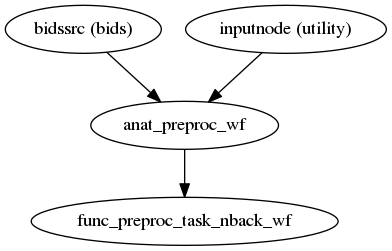
High-level view of the basic pipeline (for single-band datasets, without slice-timing information and no fieldmap acquisitions):
(Source code, png, svg, pdf)
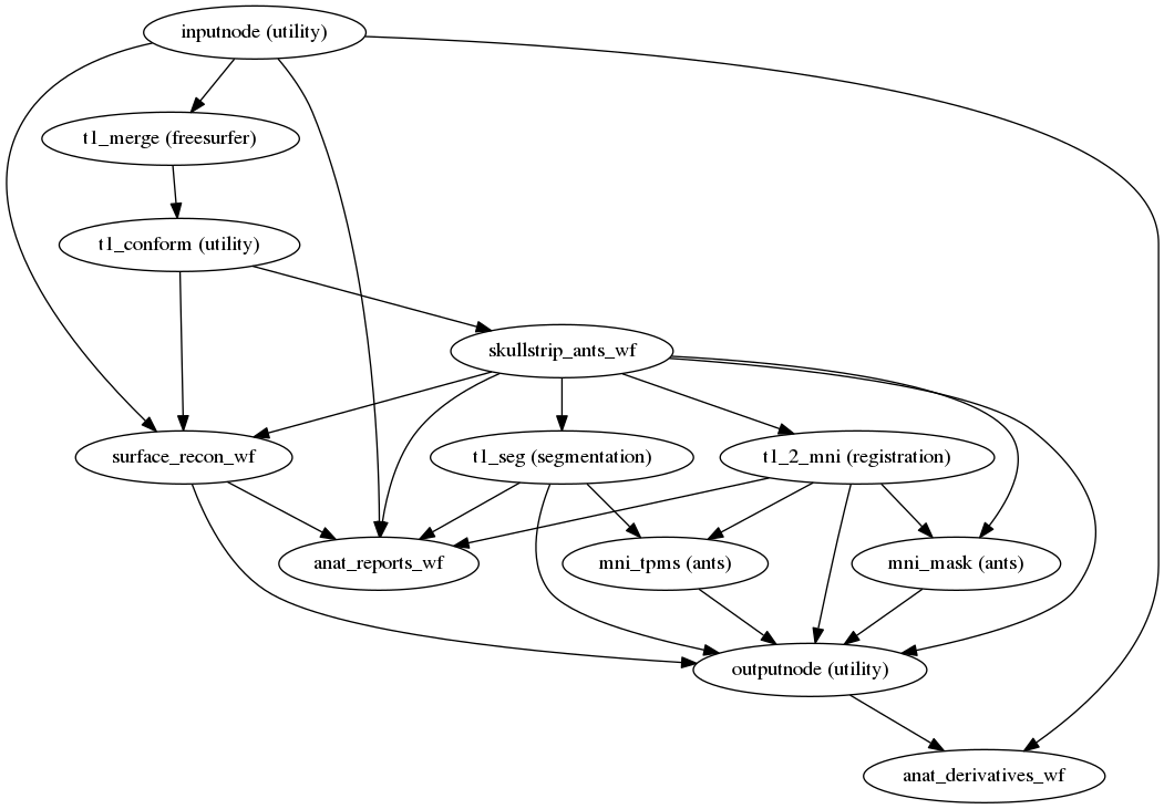
T1w/T2w preprocessing
fmriprep.workflows.anatomical.init_anat_preproc_wf
(Source code, png, svg, pdf)
This sub-workflow finds the skull stripping mask and the white matter/gray matter/cerebrospinal fluid segments and finds a non-linear warp to the MNI space.

Brain extraction (ANTs).

Brain tissue segmentation (FAST).

Animation showing T1w to MNI normalization (ANTs)
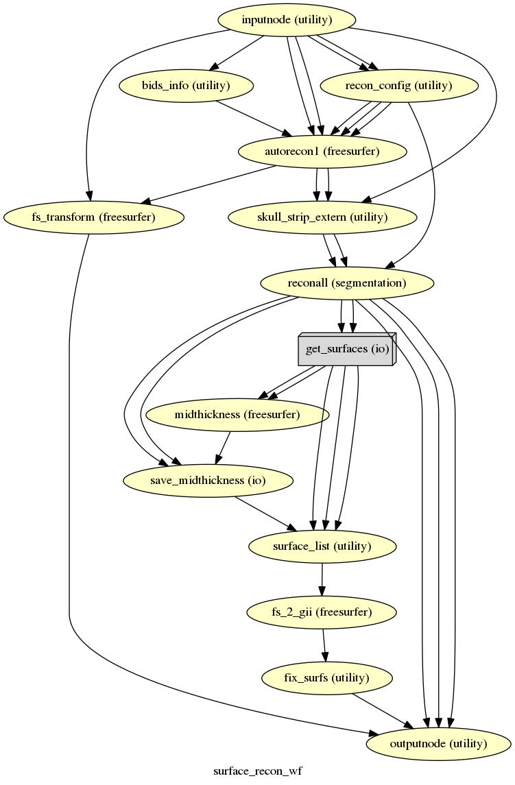
Surface preprocessing
fmriprep.workflows.anatomical.init_surface_recon_wf

(Source code, png, svg, pdf)
fmriprep uses FreeSurfer to reconstruct surfaces from T1w/T2w
structural images.
If enabled, several steps in the fmriprep pipeline are added or replaced.
All surface preprocessing may be disabled with the --no-freesurfer flag.
If FreeSurfer reconstruction is performed, the reconstructed subject is placed in
<output dir>/freesurfer/sub-<subject_label>/ (see FreeSurfer Derivatives).
Surface reconstruction is performed in three phases.
The first phase initializes the subject with T1w and T2w (if available)
structural images and performs basic reconstruction (autorecon1) with the
exception of skull-stripping.
For example, a subject with only one session with T1w and T2w images
would be processed by the following command:
$ recon-all -sd <output dir>/freesurfer -subjid sub-<subject_label> \
    -i <bids-root>/sub-<subject_label>/anat/sub-<subject_label>_T1w.nii.gz \
    -T2 <bids-root>/sub-<subject_label>/anat/sub-<subject_label>_T2w.nii.gz \
    -autorecon1 \
    -noskullstrip
The second phase imports the brainmask calculated in the T1w/T2w preprocessing sub-workflow. The final phase resumes reconstruction, using the T2w image to assist in finding the pial surface, if available:
$ recon-all -sd <output dir>/freesurfer -subjid sub-<subject_label> \
    -all -T2pial
Reconstructed white and pial surfaces are included in the report.

Surface reconstruction (FreeSurfer)
If T1w voxel sizes are less than 1mm in all dimensions (rounding to nearest
.1mm), submillimeter reconstruction is used, unless disabled with
--no-submm-recon.
In order to bypass reconstruction in fmriprep, place existing reconstructed
subjects in <output dir>/freesurfer prior to the run.
fmriprep will perform any missing recon-all steps, but will not perform
any steps whose outputs already exist.
lh.midthickness and rh.midthickness surfaces are created in the subject
surf/ directory, corresponding to the surface half-way between the gray/white
boundary and the pial surface.
The smoothwm, midthickness, pial and inflated surfaces are also
converted to GIFTI format and adjusted to be compatible with multiple software
packages, including FreeSurfer and the Connectome Workbench.
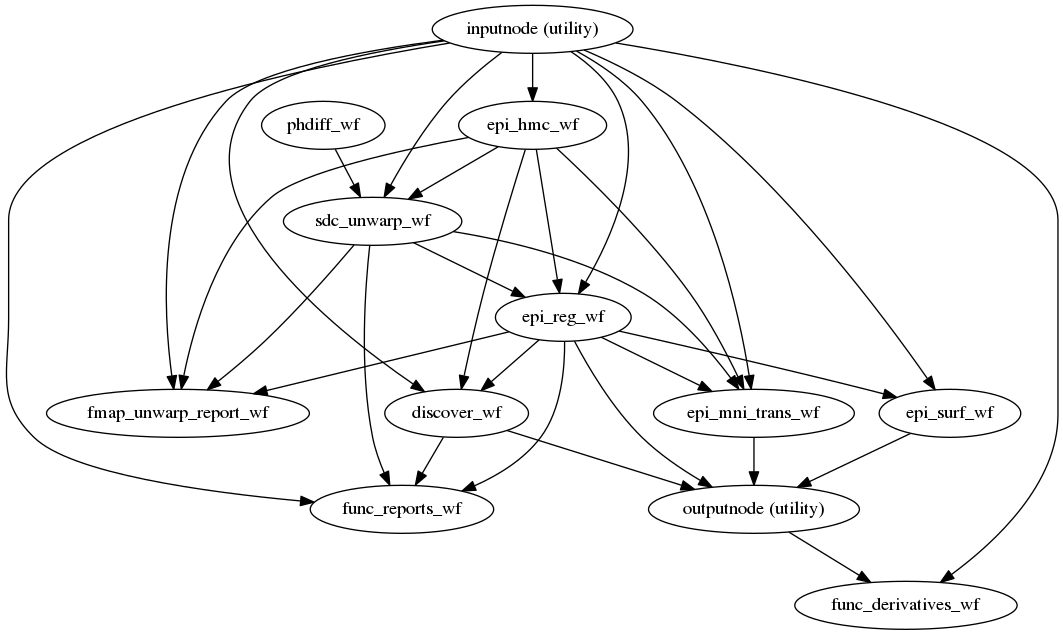
BOLD preprocessing
fmriprep.workflows.epi.init_func_preproc_wf
(Source code, png, svg, pdf)
Preprocessing of BOLD files is split into multiple sub-workflows decribed below.
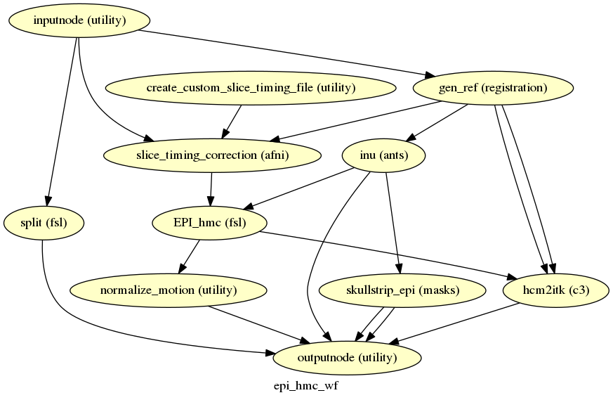
Head-motion estimation and slice time correction
fmriprep.workflows.epi.init_epi_hmc_wf

(Source code, png, svg, pdf)
This workflow performs slice time
correction (if SliceTiming field is present in the input dataset metadata), head
motion estimation, and skullstripping.
Slice time correction is performed
using AFNI 3dTShift. All slices are realigned in time to the middle of each
TR. Slice time correction can be disabled with --ignore slicetiming command
line argument.
FSL MCFLIRT is used to estimate motion transformations using an automatically estimated reference scan. If T1-saturation effects (“dummy scans” or non-steady state volumes) are detected they are used as reference due to their superior tissue contrast. Otherwise a median of motion corrected subset of volumes is used.
Skullstripping of the reference image is performed using Nilearn.

Brain extraction (BET).
Susceptibility Distortion Correction (SDC)

Applying field correction warp using ANTs.
One of the major problems that affects EPI data is the spatial distortion caused by the inhomogeneity of the field inside the scanner. The are four broad families of methodologies for mapping the field:
- Phase-difference techniques: to estimate the fieldmap, these methods measure the phase evolution in time between two close GRE acquisitions. Corresponds to the sections 8.9.1 and 8.9.2 of the BIDS specification.
- Direct field-mapping: some sequences (such as SE) are able to measure the fieldmap directly. Corresponds to section 8.9.3 of BIDS.
- Blip-up/blip-down or phase encoding polarity: pepolar: techniques acquire at least two images distorted due to the inhomogeneity of the field, but varying in PE direction. Corresponds to 8.9.4 of BIDS.
- Point-spread function acquisition: Not supported by FMRIPREP.
Once the field-map is estimated, the distortion can be accounted for. Fieldmap processing in FMRIPREP is structured as follows:
- Identifying field-mapping resources: the input BIDS dataset is queried to find the available field-mapping techniques and the appropriate processing workflows are set-up (applies to phase-difference and direct field-mapping techniques).
- Fieldmap estimation: all the estimation workflows produce a displacement field ready to be used in the correction step (applies to phase-difference and direct field-mapping techniques).
- Unwarping EPIs: the correction step is applied (for phase encoding polarity this step also involves distortion correction displacement field estimation).
If the dataset metadata indicate tha more than one field map acquisition is
IntendedFor (see BIDS Specification section 8.9) the following priority will
be used:
- Blip-up/blip-down
- Direct field-mapping
- Phase-difference techniques
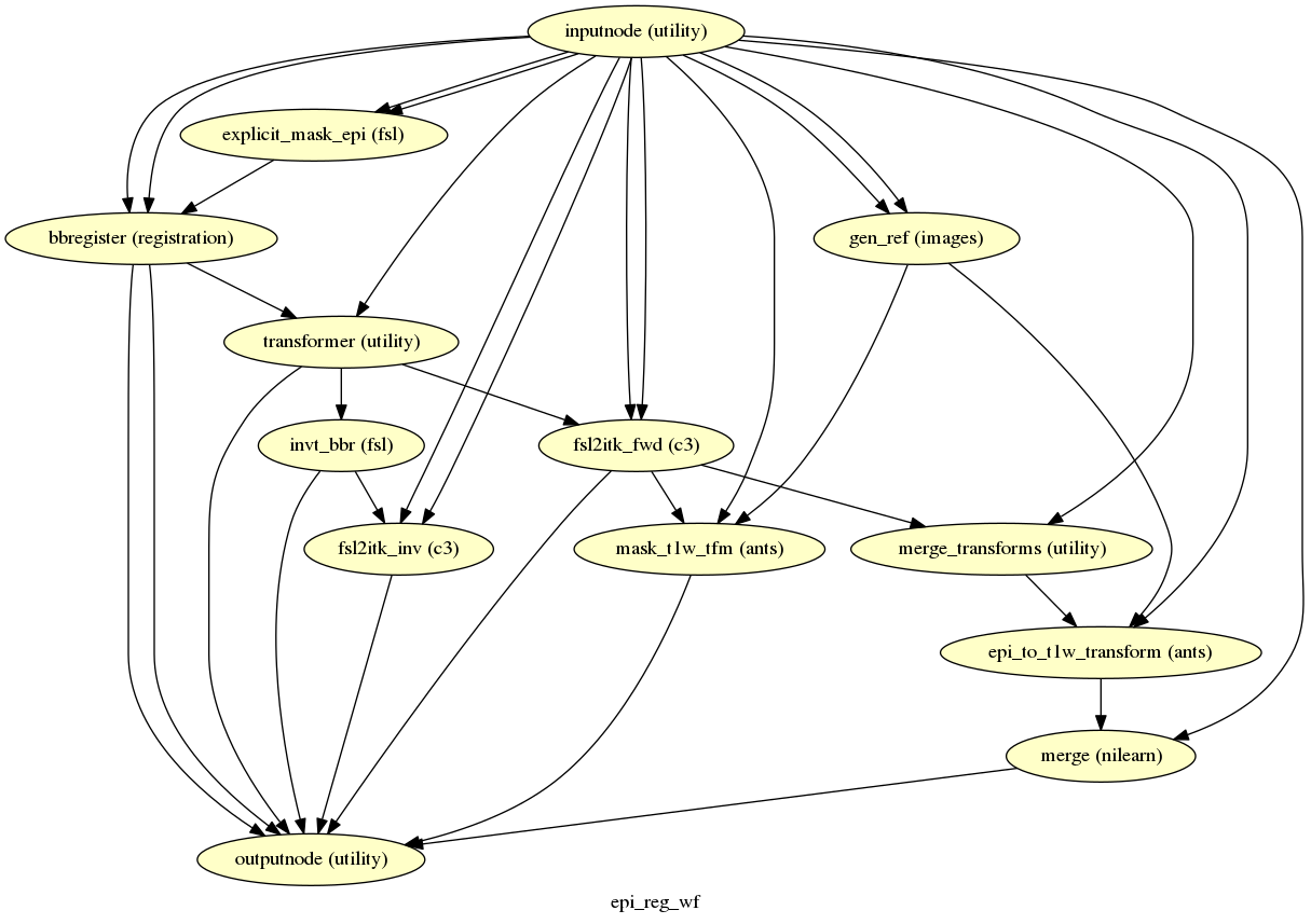
EPI to T1w registration
fmriprep.workflows.epi.init_epi_reg_wf
(Source code, png, svg, pdf)
The reference EPI image of each run is aligned by the bbregister routine to the
reconstructed subject using
the gray/white matter boundary (FreeSurfer’s ?h.white surfaces).

Animation showing EPI to T1w registration (FreeSurfer bbregister)
If FreeSurfer processing is disabled, FLIRT is performed with the BBR cost function, using the FAST segmentation to establish the gray/white matter boundary.
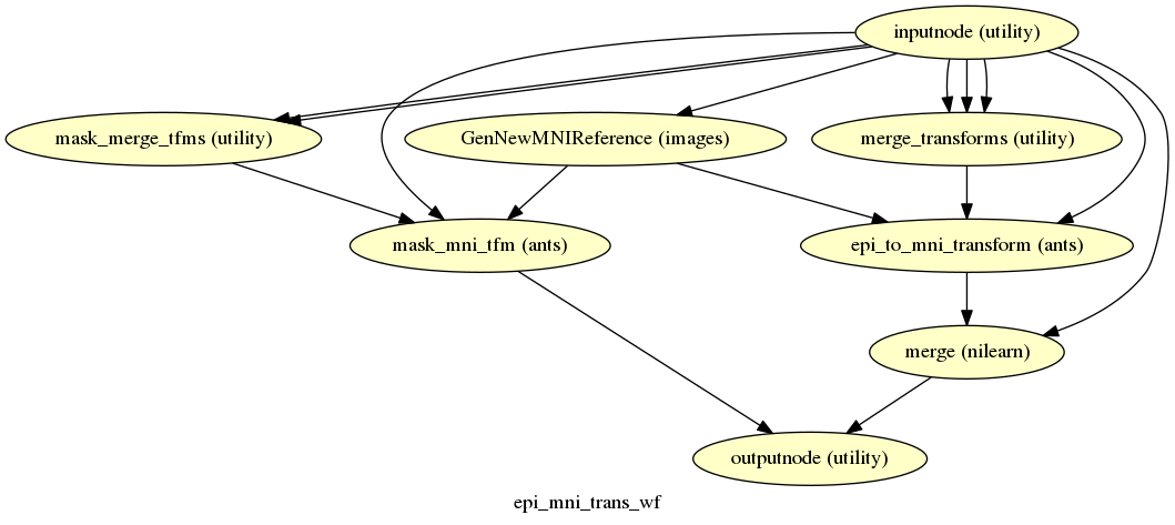
EPI to MNI transformation
fmriprep.workflows.epi.init_epi_mni_trans_wf
(Source code, png, svg, pdf)
This sub-workflow uses the transform from Head-motion estimation and slice time correction, Susceptibility Distortion Correction (SDC) (if fieldmaps are available), EPI to T1w registration, and a T1w-to-MNI transform from T1w/T2w preprocessing to map the EPI image to standardized MNI space. It also maps the T1w-based mask to MNI space.
Transforms are concatenated and applied all at once, with one interpolation (Lanczos) step, so as little information is lost as possible.
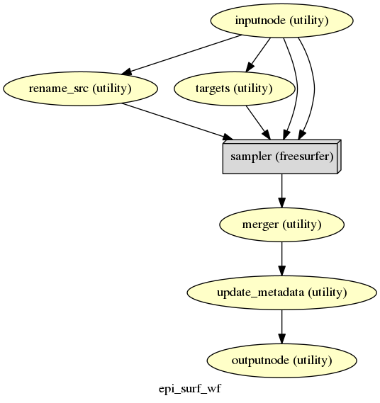
EPI sampled to FreeSurfer surfaces
fmriprep.workflows.epi.init_epi_surf_wf

(Source code, png, svg, pdf)
If FreeSurfer processing is enabled, the motion-corrected functional series (after single shot resampling to T1w space) is sampled to the surface by averaging across the cortical ribbon. Specifically, at each vertex, the segment normal to the white-matter surface, extending to the pial surface, is sampled at 6 intervals and averaged.
Surfaces are generated for the “subject native” surface, as well as transformed to the
fsaverage template space.
All surface outputs are in GIFTI format.
Confounds estimation
fmriprep.workflows.confounds.init_discover_wf
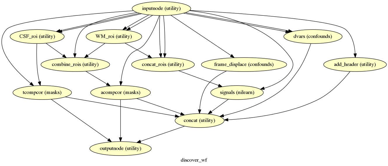
(Source code, png, svg, pdf)
Given a motion-corrected fMRI, a brain mask, MCFLIRT movement parameters and a segmentation, the discover_wf sub-workflow calculates potential confounds per volume.
Calculated confounds include the mean global signal, mean tissue class signal, tCompCor, aCompCor, Framewise Displacement, 6 motion parameters and DVARS.
Reports
FMRIPREP outputs summary reports, outputted to <output dir>/fmriprep/sub-<subject_label>.html.
These reports provide a quick way to make visual inspection of the results easy.
Each report is self contained and thus can be easily shared with collaborators (for example via email).
View a sample report.
Derivatives
There are additional files, called “Derivatives”, outputted to <output dir>/fmriprep/sub-<subject_label>/.
See the BIDS Derivatives spec for more information.
Derivatives related to t1w files are in the anat subfolder:
*T1w_brainmask.nii.gzBrain mask derived using ANTS or AFNI, depending on the command flag--skull-strip-ants*T1w_space-MNI152NLin2009cAsym_brainmask.nii.gzSame as above, but in MNI space.*T1w_dtissue.nii.gzTissue class map derived using FAST.*T1w_preproc.nii.gzBias field corrected t1w file, using ANTS’ N4BiasFieldCorrection*T1w_smoothwm.[LR].surf.giiSmoothed GrayWhite surfaces*T1w_pial.[LR].surf.giiPial surfaces*T1w_midthickness.[LR].surf.giiMidThickness surfaces*T1w_inflated.[LR].surf.giiFreeSurfer inflated surfaces for visualization*T1w_space-MNI152NLin2009cAsym_preproc.nii.gzSame as above, but in MNI space*T1w_space-MNI152NLin2009cAsym_class-CSF_probtissue.nii.gz*T1w_space-MNI152NLin2009cAsym_class-GM_probtissue.nii.gz*T1w_space-MNI152NLin2009cAsym_class-WM_probtissue.nii.gzProbability tissue maps, transformed into MNI space*T1w_target-MNI152NLin2009cAsym_warp.h5Composite (warp and affine) transform to transform t1w into MNI space
Derivatives related to EPI files are in the func subfolder.
*bold_confounds.tsvA tab-separated value file with one column per calculated confound and one row per timepoint/volume
Volumetric output spaces include T1w and MNI152NLin2009cAsym (default).
*bold_space-<space>_brainmask.nii.gzBrain mask for EPI files, calculated by nilearn on the average EPI volume, post-motion correction*bold_space-<space>_preproc.nii.gzMotion-corrected (using MCFLIRT for estimation and ANTs for interpolation) EPI file
Surface output spaces include fsnative (full density subject-specific mesh),
fsaverage and the down-sampled meshes fsaverage6 (41k vertices) and
fsaverage5 (10k vertices, default).
*bold_space-<space>.[LR].func.giiMotion-corrected EPI file sampled to surface<space>
FreeSurfer Derivatives
A FreeSurfer subjects directory is created in <output dir>/freesurfer.
freesurfer/
    fsaverage{,5,6}/
        mri/
        surf/
        ...
    sub-<subject_label>/
        mri/
        surf/
        ...
    ...
Copies of the fsaverage subjects distributed with the running version of
FreeSurfer are copied into this subjects directory, if any functional data are
sampled to those subject spaces.